Aree di Ricerca:
Advanced Materials -Chemistry for Cultural Heritage – Chemistry for Life Sciences – Energy – Green Chemistry
Parole chiave:
Morphologic characterization, Optical Microscopy, Atomic Force Microscopy, Confocal Microscopy
Contact persons:
Marinella Striccoli – marinella.striccoli@cnr.it
+390805442027
Chiara Ingrosso – chiara.ingrosso@cnr.it
+390805442027
Altre informazioni:
The laboratory deals with the morphological and optical characterization of nanostructured, nanocomposites and thin-film materials and is equipped with an atomic force microscope (AFM), a confocal microscope, an optical transmission microscope also in polarized light and a stereo microscope.
The broad range of applications available to laser confocal microscopy includes a wide variety of studies in material science as well as morphological studies of a wide spectrum of cells and tissues. Other applications include resonance energy transfer, stem cell research, photobleaching studies, total internal reflection, bioluminescent proteins etc.
The AFM allows the investigation of the morphological properties of functionalized surfaces, nanocomposite materials and 2D/3D assembly of nanoparticles and nanocrystals on substrates.
ELENCO DELLA STRUMENTAZIONE DISPONIBILE

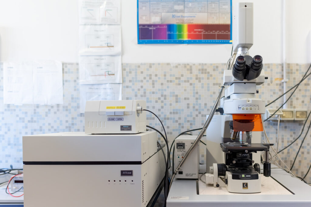
A Nikon C1 confocal system, equipped with a Nikon Eclipse E600 direct microscope and 2 laser (a diode module at 405 nm and a multiline Ar laser with emission at 457/488/514nm). Its scanning head has 3 photomultiplier tubes (PMTs) which can detect simultaneous 3-channel fluorescence in double- and triple-labelling experiments. The microscope has several objectives in air (from 5X to 40X and with a 60X in oil.) A digital camera provide bright-field and fluorescent imaging capabilities. The confocal system delivers 3D fluorescent images of samples with high resolution and contrast, allowing analysis of fluorescent labeled thick specimens without physical sectioning and 3-dimensional reconstruction (XYZ) and time-analysis (XYT).

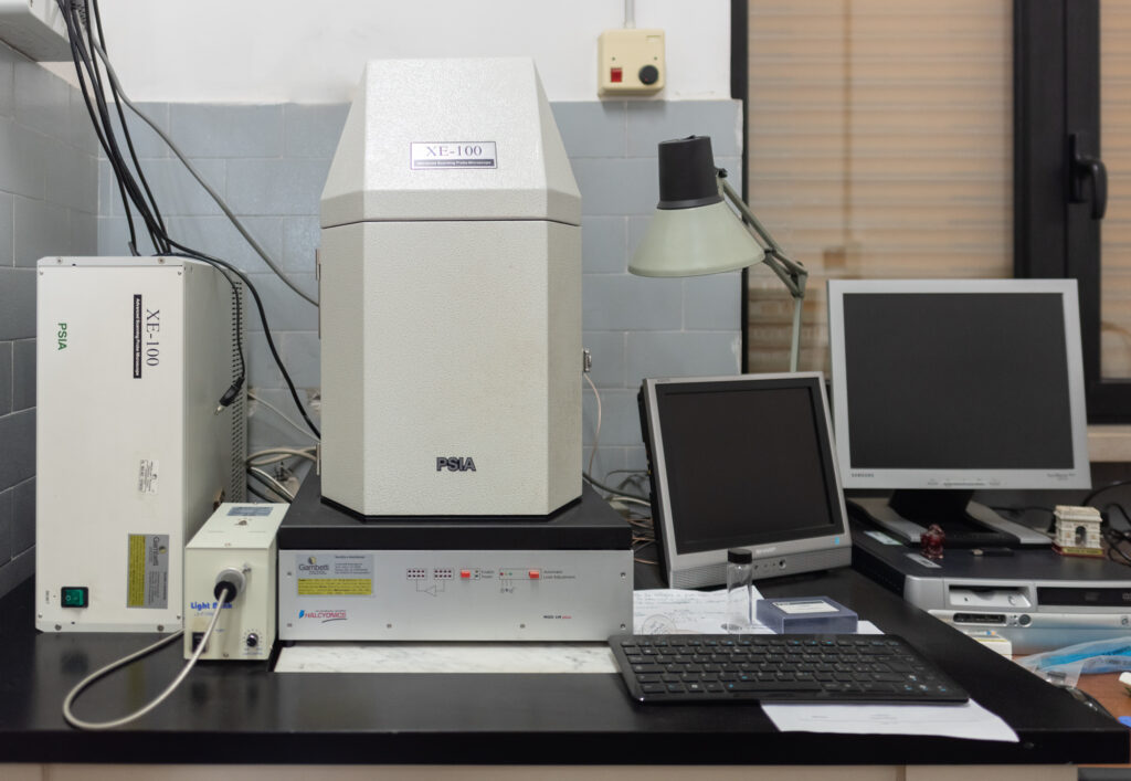
Atomic Force Microscope AFM XE 100 PSIA with decoupled XY and Z scanner, and single module flexure XY scanner with closed-loop control, working in True Non-Contact Mode and Contact mode. Scan range of XY scanners: 5 µm, 50 µm, or 100 µm, Scan range of Z scanners: 12 µm or 25 µm. Optical resolution: 1 µ.
